 | Questions |
 | Major surgery is not the only option for many patients with disc herniation. Call 800-956-6724.
| |
|
| |
| Journals |
|
 |
| | |
 |
 |
|
Low back pain is one of the most commonly encountered problems in a medical practice. Diskogenic low back pain, or pain arising from the intervertebral disks, has been estimated to be the most common cause of chronic low back pain. Historically, open surgery techniques have been used to treat diskogenic pain, and more recently minimally invasive techniques have been developed. However, "minimally invasive" has become a trite phrase that has lost its meaning and is used by different authors to describe totally different things. In the spine field, it is time to replace this language with a less ambiguous perspective focused on whether there is access trauma or not. Any procedure that is larger in the approach to the pathology than is necessary creates potential morbidity for the patient from the access itself. A tremendous advantage of all percutaneous procedures is that they are, by their nature, nontraumatic in access. Having stated the obvious, that nontraumatic access is beneficial, what remains is to determine how to optimally treat pathology with techniques and technology that clearly must be seen as restricted--restricted as to the extent of expansion of exposure at the level of the pathology, and also restricted to those practitioners with specialized expertise.
|
| |
|
Classification
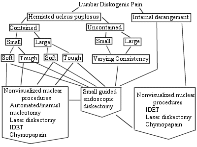
Although no universally accepted classification system has been suggested for percutaneous techniques for diskogenic pain, the following definitions are helpful. Diskogenic pain is thought to be produced by internal disk derangement and herniated nucleus pulposus (HNP) (disk herniation), both of which occur as a spectrum of pathology.
Disk herniations can be further subdivided into contained (within disk boundaries) and uncontained (outside of disk boundaries and into the spinal canal). A spectrum of herniations is seen based on size and consistency.
Intranuclear procedures (both visualized and nonvisualized) are used to treat contained HNP and internal disk derangement. Extranuclear procedures (visualized) are used to treat uncontained HNP
|
| |
 Workup Workup
History and Physical Examination
The workup for percutaneous procedures is identical to the workup of disk problems in general. A focused history and physical examination are performed.
Spinal Imaging
Imaging studies are obtained. The most useful study is magnetic resonance imaging (MRI) to directly visualize the disk pathology. Alternatively, computed tomographic (CT) myelography is helpful to visualize canal encroachment, and discography is useful to visualize the integrity of the disks and determine if they reproduce the patient's pain symptoms.
|
| |
|
Treatment
Nonvisualized Nuclear Procedures.
Although these procedures utilize different physical methods, the basic principle is the same. They seek to reduce the volume of the disk by removal of 10 to 15% of the nuclear material and thus reduce intradiskal pressure. Advocates believe that this will help to reduce back pain and nerve root compression.
The four most commonly used nonvisualized intranuclear procedures are chemonucleolysis, percutaneous automated nucleotomy, percutaneous laser disk decompression (PLLD), and intradiskal electrothermal therapy (IDET).
Chemonucleolysis
In 1963, Smith administered the enzyme chymopapain to the first patient. By 1984, 75,000 patients had been treated with this method. Unfortunately in 1999, this procedure was widely discontinued in the United States after several cases of transverse myelitis and anaphylactic deaths were reported.
Percutaneous Automated Nucleotomy
In 1975 Hijikata introduced the percutaneous manual nucleotomy, which was expanded by Onik, a radiologist, who developed and automated device (Nucleotome, Clarus Medical, Minneapolis, MN). This consists of a 2.5-mm probe that is positioned into the nuclear chamber via a standard posterolateral approach. The probe contains a cutter and a suction mechanism. The nuclear material is cut and suctioned to an outside reservoir. The reported success rates of this procedure by itself vary from 29% (Chatterjee, 1995) to 75% (Onik et al, 1990). It is a relatively simple and safe technique and major complications are very rare.
Percutaneous Laser Disk Decompression (PLDD)
Since its introduction by Ascher and Choy in 1986, the utilization of laser energy to reduce the nucleus pulposus volume has gained increasing popularity due to its small size, technical simplicity, and low incidence of complications. The effectiveness of PLDD varies from 75 to 90% (as reported by Choy) to 60 to 85% (satisfactory as reported by Maroon); but Knight demonstrated clinical benefit in only 52% with an additional 21% rate of functional improvement. Unfortunately, these studies are more anecdotal, and no high-quality controlled trials have been performed. The incidence of complications is around 1% and includes infectious diskitis, cauda equinasyndrome, bowel perforation, and nerve root damage.
Intradiskal Electothermal Therapy (IDET)
The intradiskal electrothermal treatment (IDET, Oratec Interventions INC., Menlo Park, CA) was introduced by physiatrists Jeffrey and Joel Saal. The mechanism of action of this procedure is a subject of controversy and is not well described in the literature. Complications are rare. Thomas et al (2004) reported a case in which the tip of the catheter broke off into the spinal canal and migrated to inside the dural sac, which was later removed by a wide laminectomy. Long-term data collection found that the results were less promising than previously expected. Pauza et al (2003) reported that even though 40% of their patients achieved greater than 50% relief of their pain, about 50% of the patients experience no appreciable benefit.
Visualized Intra-/Extranuclear Procedures
Small Guided Endoscopic Diskectomy
Advocates believe that this procedure can be used to treat all causes of diskogenic pain, including contained HNP, uncontained HNP, or internal derangement. Diskectomy is performed through a small working channel endoscope, passed through a tapered cannula. Direct visualization is possible, but the access is small and minimally traumatic. Decompression of the disk and the delivery of uncontained free fragments from the spinal canal are possible, without cutting the muscle, removing bone or yellow ligament, or retracting the dural sac and nerve roots (Fig 63-1). Other forms of energy such as manual or automated nucleotomy, laser, and IDET may be delivered through the working channel of the arthroscope.
|
| |
A 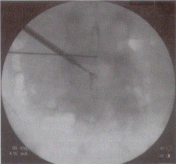 B B
C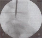
D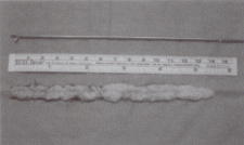
|
| Figure 63-1 (A)Axial compyted tomography (CT) postdiskogram showing location of migrated fisk fragments behind the vertebral body. (B) Anteroposterior (AP) and (C) lateral intraoperative view showing grasper placement to remove migrated fragments. (D) Disk fragments that have been removed with the micrograsper through the small endoscope. | | |
|
Outcome
Well-controlled outcome studies have not been performed for most of the above techniques at the time of this writing.
|
| |
|
Complications
Complications include infection, neurologic injury, retained pathology, continued pain, and injury to vital structures.
|
| |
|
Suggested Readings
Davis JK. Percutaneous laser discectomy. In: Cohen AR, Haines SJ, eds. Minimally Invasive Techniques in Neurosurgery: Concepts in Neurosurgery. Vol 7. Baltimore: Williams & Wilkins; 1995:254-257
Discusses laser diskectomy techniques.
Ditsworth DA. Endoscopic transforaminal disc removal and reconfiguration. Presented at the Spine Disorders 1996 annual meeting joint section (CNS/AANS) spine and peripheral nerves, 1996
One of the initial descriptions of the technique of small guided Endoscopic transforaminal disk removal.
Ditsworth DA. Endoscopic transforaminal lumbar discectomy and reconfiguration: a posterolateral approach into the spinal canal. Surg Neurol 1998;49:588-598
Describes indications, contraindications, technique, risks, and results for small guided Endoscopic diskectomy.
Ditsworth, DA. IDET and PED: Benefits of Combination Treatment. Presented at the Spine Disorders 2000 annual meeting joint section (CNS/AANS) spine and peripheral nerves, 2000.
Combines IDET and the small endoscope; adding visualization provides additional benefit.
Hijikata S. Percutaneous nucleotomy: a new concept technique and 12 years experience. Clin Orthop Relat Res 1989;238:9-23
Discusses percutaneous nucleotomy.
Javid MJ. Chemonucleolysis. In: Cohen AR, Haines SJ, eds. Minimally Invasive Techniques in Neurosurgery: Concepts in neurosurgery. Vol 7. Baltimore: Williams & Wilkins; 1995:240-246
Describes the technique of chemonucleolysis, including risks and complications.
Onik G. Mooney V, Maroon JC, et al. Automated percutaneous discectomy: a prospective multi-institutional study. Neurosurgery 1990;26:228-233
Give results of basic nonvisualized diskectomy via nucleotome gathered from numerous institutions.
Smith L. Enzyme dissolution of the nucleus pulposus in humans. JAMA 1964;136:376-379
A foundational article regarding enzyme dissolution of disk.
|
| |
|
© 1995-2006 SPINEONLINE.COM
Any questions about this site, mail to Webmaster.
Please read our policy before using this site. |
|

